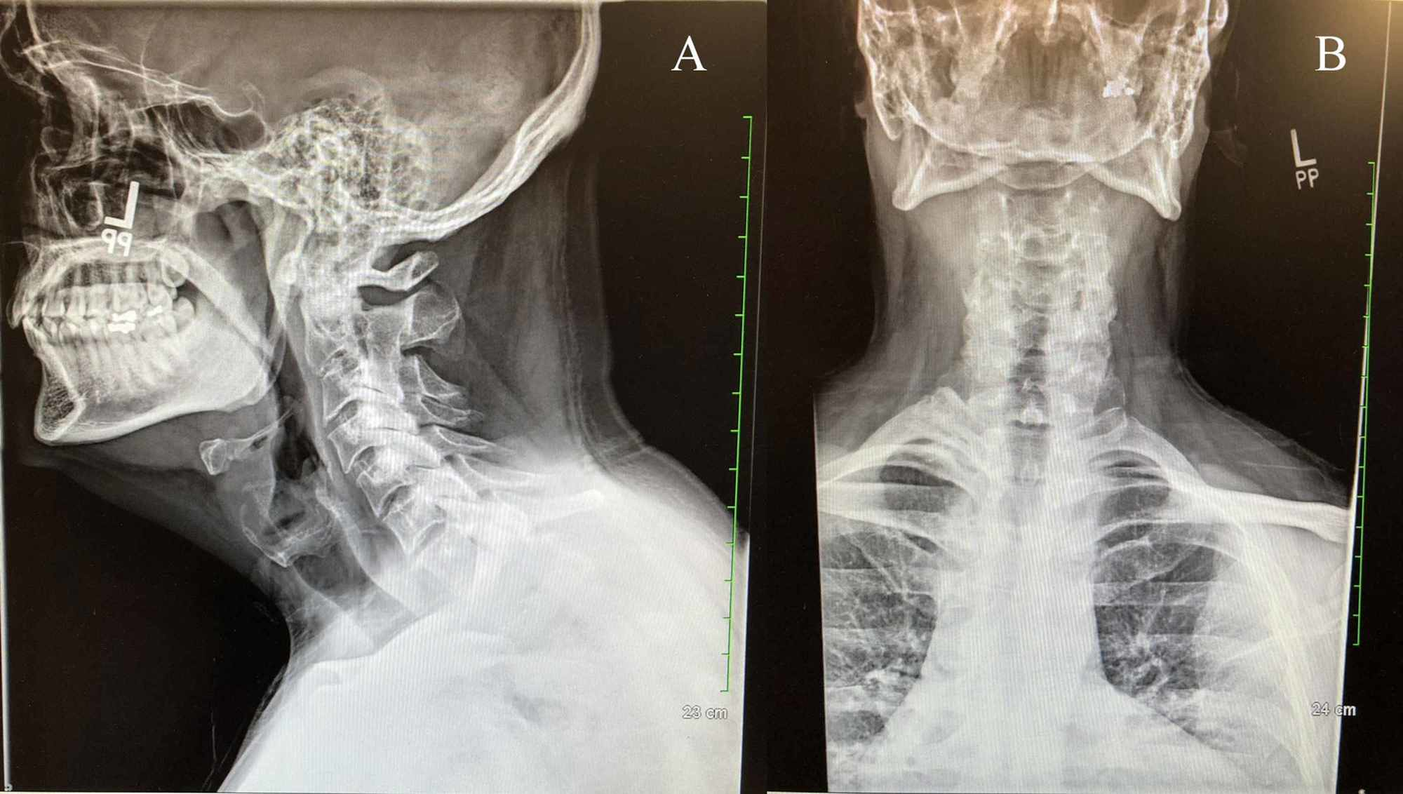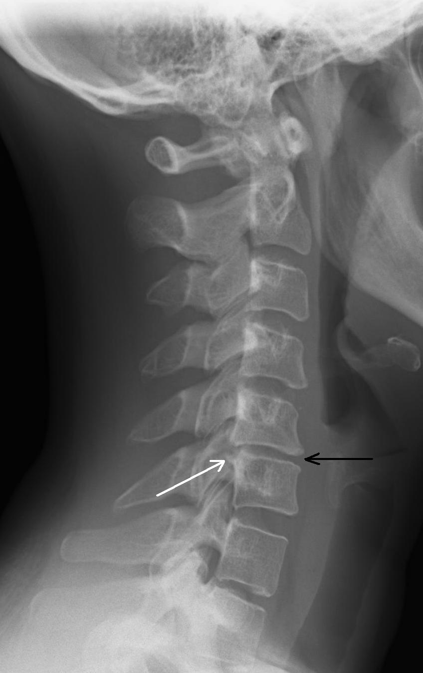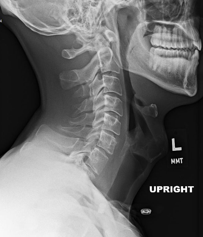

The bodies and spinous processes of C-2 to C-7 are fully visualized, and the intervertebral disk spaces and prevertebral soft tissues can be adequately evaluated. This projection suffices to demonstrate most traumatic conditions of the cervical spine, including injuries involving the anterior and posterior arches of C-l the odontoid process, which is seen in profile and the anterior atlantal-dens interval. The single most valuable projection in these instances is the lateral view, which may be obtained in the standard fashion or with the patient supine, depending on their condition. Frequently the patient is unconscious, there are associated injuries, and unnecessary movement risks damage to the cervical cord.

Radiographic examination of a patient with cervical spine trauma may be difficult and is usually limited to one or two projections.

Extending laterally from the pedicle on each side is a transverse process, and in the posterior portion, a spinous process extends from the junction of the laminae in the midline. Extending caudad and cephalad from the junction of the pedicle and lamina on each side are superior and inferior articular processes, which form the apophyseal joints between the successive vertebrae. The vertebrae C-3 to C-7 exhibit identical anatomic features and are more uniform in appearance, consisting of a vertebral body and a posterior neural arch, including the right and left pedicles and laminae, which together with the posterior aspect of the body enclose the spinal canal. The second vertebra, C-2 or axis, is a more complex structure whose distinguishing feature is the odontoid process, also known as the "dens" (tooth), projecting cephalad from the anterior surface of the body. The atlas has no body its main weight-bearing structures are the lateral masses, also called articular pillars. The first cervical vertebra, C-l or atlas, is a bony ring consisting of anterior and posterior arches connected by two lateral masses. Structurally, the first and second cervical vertebrae possess anatomic features distinct from those of the remaining five cervical vertebrae. Oblique projections of the cervical spine are not routinely obtained, although they may be called for to help visualize obscure fractures of the neural arch and abnormalities of the neural foramina and apophyseal joints. If you'd like to comment on or contribute to this series, please e-mail most common routine cervical projections are the anteroposterior (AP), AP open mouth, and lateral.
Cervical spine x ray series#
This article is the 14th in our series of white papers on radiologic patient positioning techniques for x-ray examinations. Neck injury caused by a sudden jerking of the head is commonly called whiplash.Radiographic positioning techniques for the cervical spine By Dr. If your neck is dislocated or fractured, your spinal cord may also be damaged. This is especially true with falls, car accidents, and sports, where the muscles and ligaments of the neck are forced to move outside their normal range.

The neck is particularly vulnerable to injury. Your doctor may request a neck X-ray if you have a neck injury or pain, or persistent numbness, pain, or weakness in your arms.


 0 kommentar(er)
